Biomedical Research and Mentoring Program
Cincinnati Children's Hospital Medical Center
January 2015 - May 2015
|
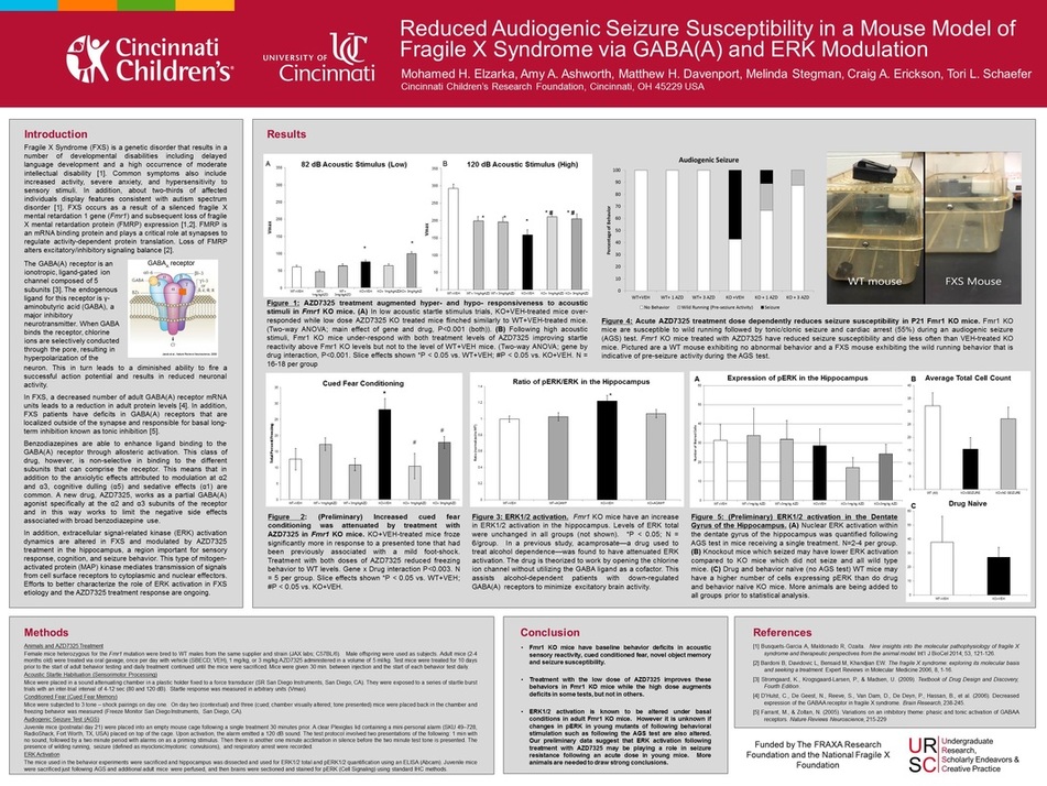
In the spring semester of 2015, I participated in the University Honors Biomedical Research and Mentoring Program, which pairs honors students with faculty researcher mentors at the University of Cincinnati College of Medicine and Cincinnati Children's Hospital Medical Center. Through the program, I had the opportunity to research in the Developmental Disabilities Translational Research Center, working with and learning from Dr. Tori Schaefer. Dr. Schaefer's research is focused on developing targeted drug treatments for developmental disabilities like Fragile X Syndrome, Angelman Syndrome, and Autism Spectrum Disorders using mouse models for these diseases. During my time in the lab, the primary research focus was on Fragile X Syndrome--a genetic disorder that is resultant of a gene mutation on the X Chromosome that causes increased excitation in the brain, along with a host of other physical and behavioral phenotypes. Using a repurposed drug designed originally to combat Generalized Anxiety Disorder, the lab studied the effects of enhancing the activation of the inhibitory GABA(A) receptor through allosteric binding. My specific project while in the lab was focused on analyzing the effects of this drug with regard to audiogenic seizure susceptibility and hippocampal activation. At the end of the semester, I was able to present this research at the Undergraduate Research Conference hosted by the Office of Undergraduate Research, Scholarly Endeavors, and Creative Practice.
I initially decided to pursue a spot in the Biomedical RaMP because of the great experiences I have had in previous research involvements. Prior to joining the program, I was involved with the High School Senior Summer Internship at Cincinnati Children's, the Excellence in Science Education and Learning (ExSEL) Program funded by the Howard Hughes Medical Institute, and a brief foray into research with the UC Department of Neurosurgery. From all of these experiences, I developed a strong interest in and passion for research, specifically in neuroscience. The Schaefer lab was a great fit because of the translational nature of the work--which relates to my interest in a combined MD/PhD degree--and because the lab's research explores the interplay between neuroscience and genetics, two pivotal areas of medicine moving into the future that are of great interest to me. Thankfully, my time in the lab was engaging and insightful, and the experience that I had helped develop my scientific understanding and technical skills. The experience was a meaningful one because of its ability to truly open my eyes to what a career in academic medical research entails. I learned that research is a career of dedication and perseverance, and that patience and an investment of large amounts of time are extremely necessary for success. I also learned that it is vital to balance a strong foundation in scientific study with the technical skills needed to transfer knowledge into action. Moving forward, I will take into strong consideration that the labor intensiveness of research may pose an obstacle to a career as both a physician and scientist. I do not yet know what path in medicine will be the best fit for me, but I hope that as I continue to refine my technical skills in research with more lab experience and to strive for success in my classwork, I will find my way. The embedded proposal for this Honors Experience details the important strides--with regard to both an increased working knowledge of scientific principles and technical procedures and an established course of personal growth--that I sought to take through the completion of this program.
|
This presentation was given to fellow students and their mentors as part of the dissemination process for this Honors Experience. This presentation, in contrast to my formal research poster presentation at the Undergraduate Research Conference, is more focused on the reflective learning that I was able to take away from my time in the Erickson-Wink Lab rather than some of the more detailed scientific questions that I was investigating.
|

As part of my experience in the Biomedical Research and Mentoring Program, I was fortunate enough to have the opportunity to present some of my findings at the Office of Undergraduate Research, Scholarly Endeavors and Creative Practice's 2015 Undergraduate Research Conference. My research focused on Fragile X Syndrome, a genetic disease that is the largest single cause of intellectual disability that can be inherited. The disease is often associated with over-excitation in the brain, resulting in behaviors like extreme anxiety, increased activity, and hypersensitivity to sensory stimuli. In addition, the over-excitation is responsible for the fact that 15% of males and 5% of females with Fragile X Syndrome suffer from seizures. Using a mouse model for the disease, the lab studied whether the excitatory activity associated with Fragile X Syndrome and the resultant phenotypes could be improved through the use of a drug modulator of the inhibitory neurotransmitter GABA. By increasing the efficacy of binding at the GABA receptor, the drug is theorized to result in the improved outcomes that mice have in tests meant to measure anxiety, responses to sensory stimuli, and seizure susceptibility.
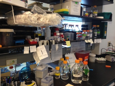
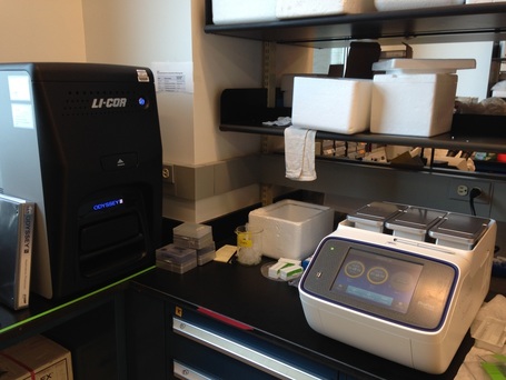
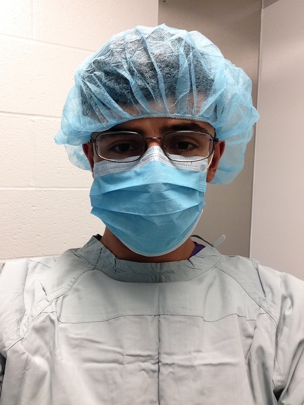
With time spent both in the wet lab at the bench and in the behavioral testing, animal handling, and necropsy facilities, my research experience this semester has been multidimensional.
|
|
|
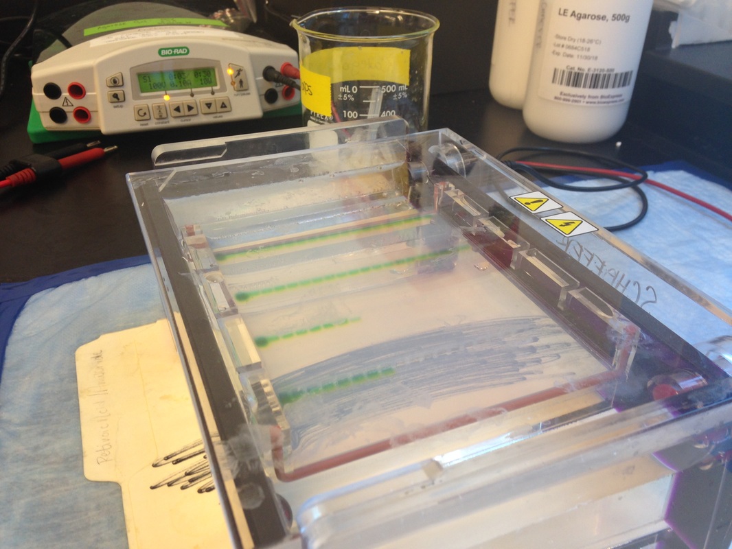
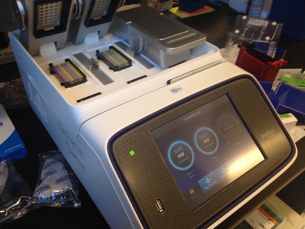
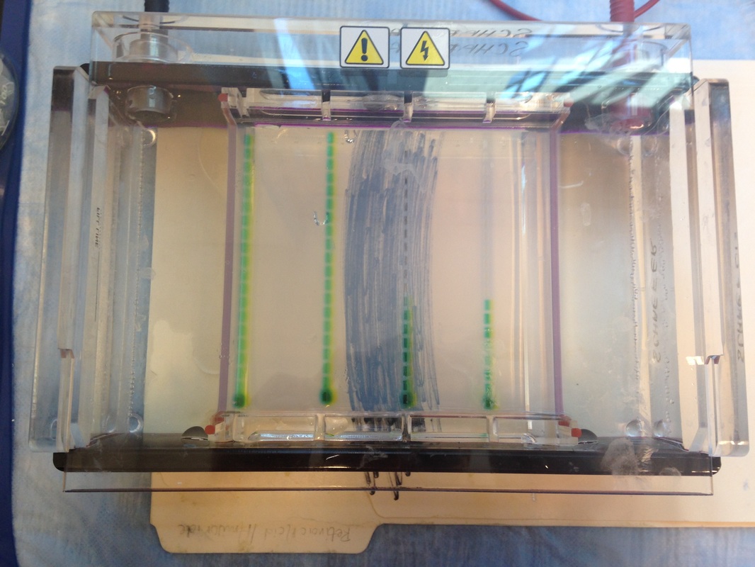

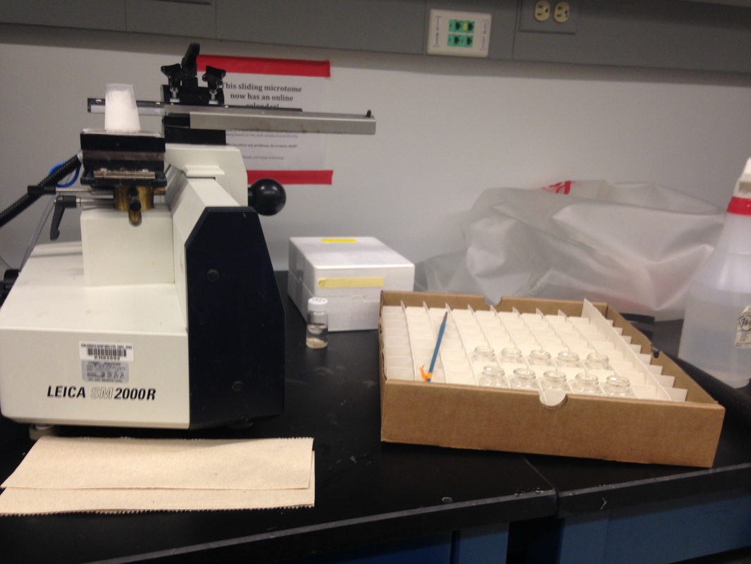
Knowing the genotype of all mice used in a study is a vital component of any genetic research. In order to determine the genotype of a mouse, a sample of tissue like an ear clip or portion of tail must be obtained. This sample must then be digested by detergents that break open the cell and expose the DNA. After the DNA is replicated through the process of polymerase chain reaction, it can be run through gel electrophoresis to produce banding indicative of different genotypes.
|
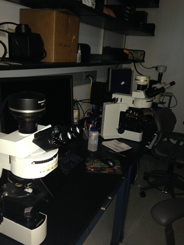
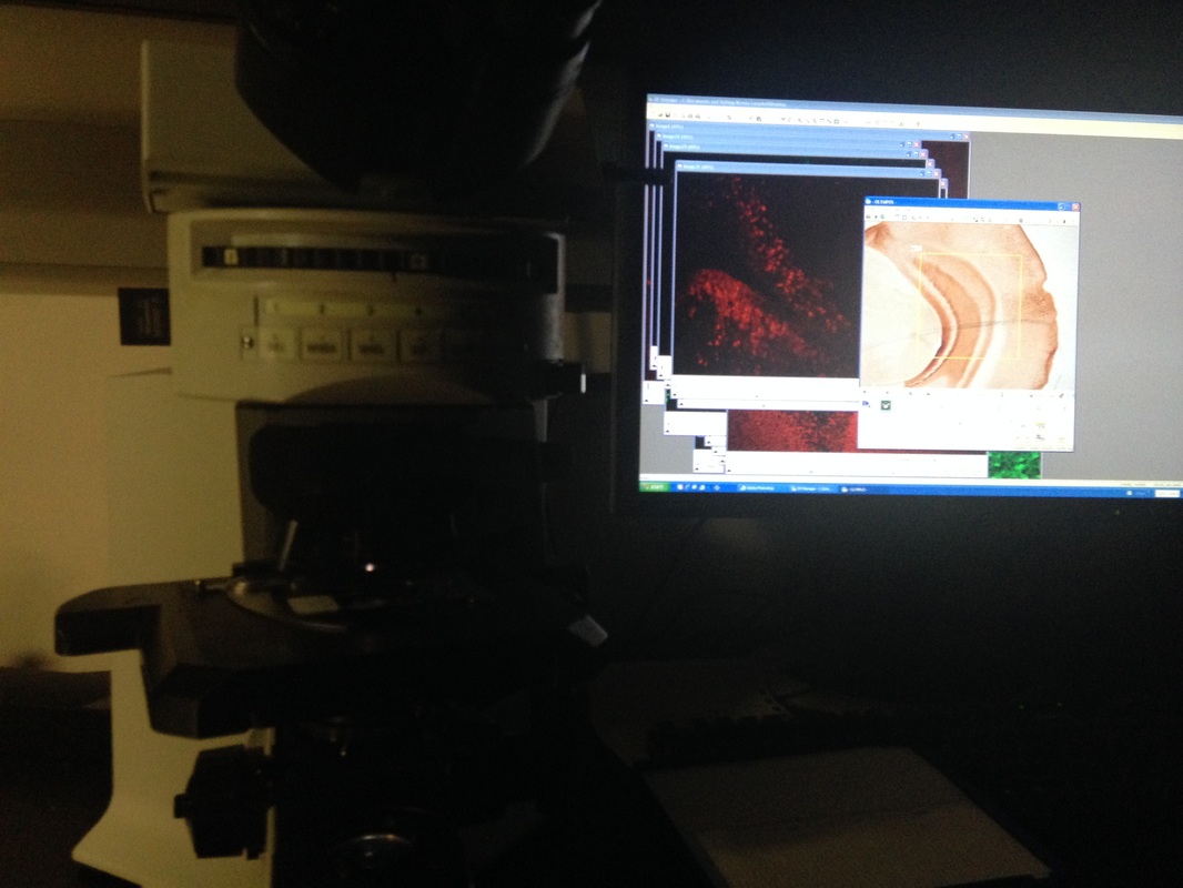
The microscopy room is home to the microscopes and imaging software that are used to image the brain sections that have been stained with immunohistochemistry (IHC).
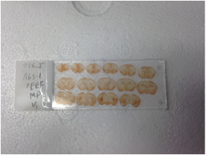
For the audiogenic seizure project on which I worked, wild-type and disease-state mice with different treatment dosages were exposed to loud tones that induced seizure activity in some mice. When the brains of the mice were collected and sliced, these brains could be stained via IHC to localize ERK activation that is indicative of cell activity. Pictured below is an image of the dentate gyrus of the hippocampus, which had lessened activity in those knockout mice that did seize as compared with those that did not. 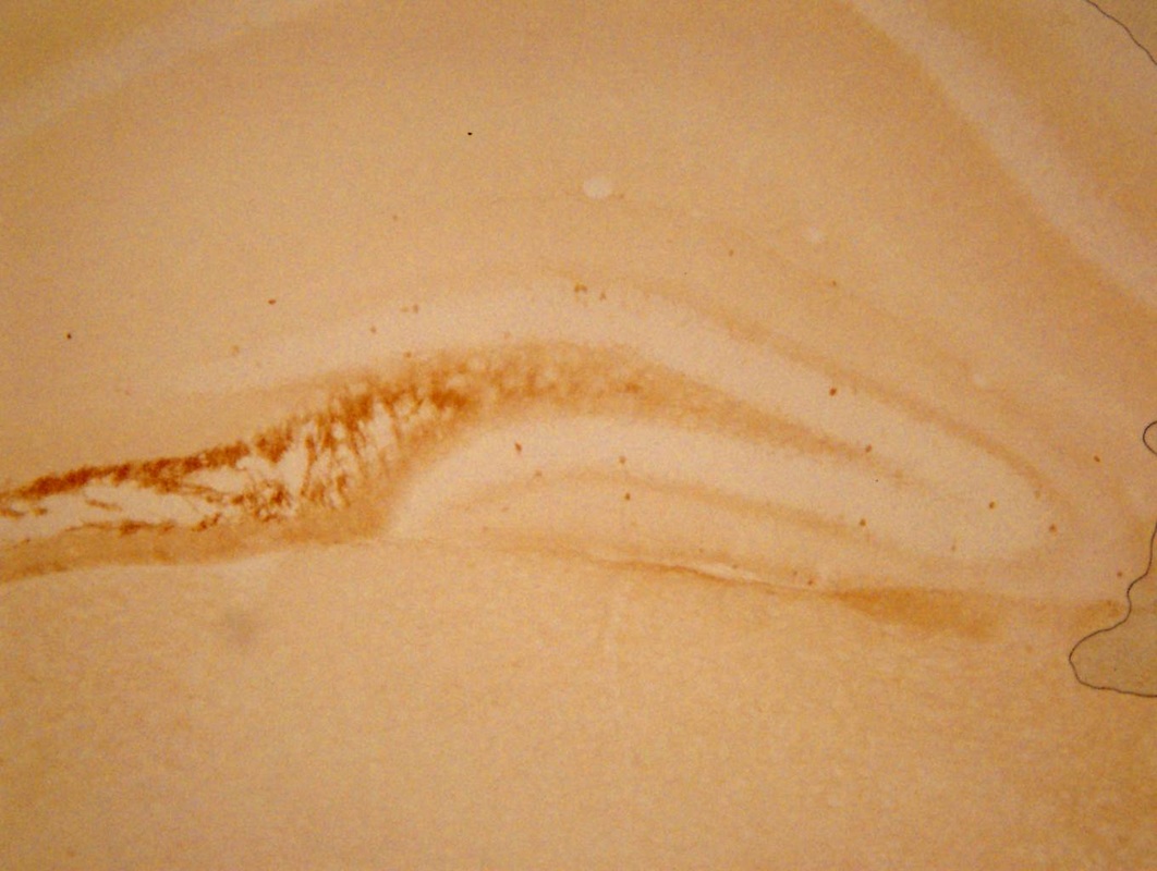
|
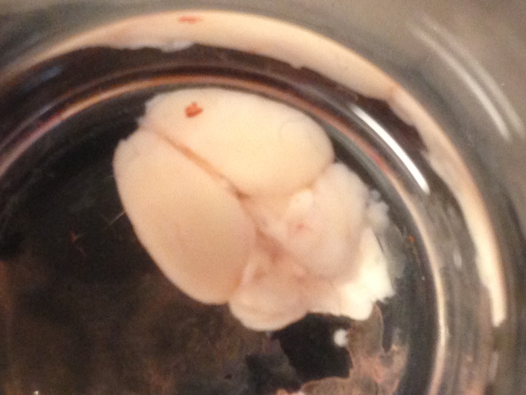
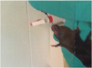
A perfusion is by definition the process of diffusing a liquid through something. In the case of mice, perfusing the fixative paraformaldehyde through the blood vessel vasculature allows for the fixation--or preservation from decay--of the mouse brain. After fixation, the obtained brains can be sectioned, stained, and then imaged to determine differential activations of specific brain regions.
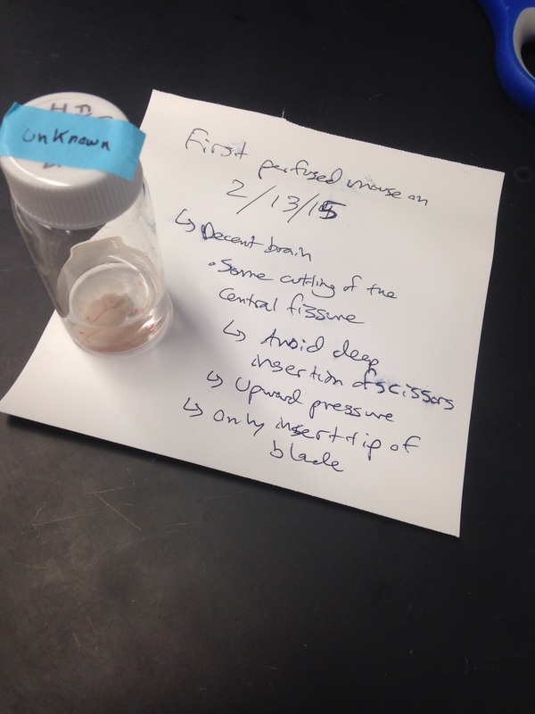
My own experience with perfusion was positive. While I never reached the level of expertise that other lab members had after year of practice, I was able to effectively perfuse several mice for the collection of their brains. Pictured above is my first perfused brain, along with notes indicating areas of improvement in the perfusion technique.
|
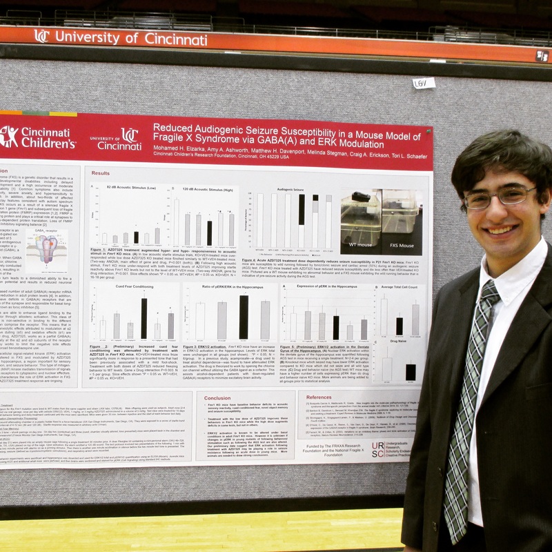
My presentation at the Undergraduate Research Conference was a rewarding experience.
I would like to thank Dr. Tori Schaefer, Dr. Craig Erickson, and the rest of the lab team for facilitating an engaging and immersive semester.
|
Embedded is my reflective essay for this experience, which details how much of an insightful, challenging, and beneficial experience my involvement in the lab this semester was. While I was not able to accomplish all that I had set out to, I did achieve many of my goals and learned a great deal.
|
