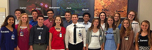
High School Senior Summer Internship
Cincinnati Children's Hospital Medical Center
June 2014 - August 2014

In the summer between graduating high school and beginning life as a Bearcat, I had the opportunity to work with Dr. Douglas Rose in the Neurology Department of Cincinnati Children's Hospital. I worked with Dr. Rose as part of a paid internship program through Cincinnati Children's, called the High School Senior Summer Internship. The program, designed for 17 of Cincinnati's highest-achieving graduating seniors, paired students with mentors in one of 15 pediatric clinical specialties. My experience with Dr. Rose was wonderful because it allowed me to shadow Dr. Rose and resident physicians in the department as they made their way on patient rounds and to engage in the research efforts being pursued by Dr. Rose with regard to locating the epileptic focus of seizures.
My experience with Dr. Rose and the Neurology Department was a very insightful and engaging one. As part of my internship, I was able to shadow resident and attending physicians on their clinical rounds. This was my first true experience seeing the doctor-patient relationship up close, and it was very rewarding to witness the personalized attention that each doctor gave to those in their care. It was also very interesting to witness how collaborative all of the doctors were with one another, both in addressing patient needs and in determining appropriate treatment methods. This cooperation definitely had a positive impact for patients, as the constant questioning and evaluating, and the different perspective, that each doctor brought to the table helped construct a very carefully thought out plan for care. From the patient rounds, I was also made aware of more diagnostic tools and of diseases with which I had previously been unfamiliar. In addition, I was able to shadow physicians in the Divisions of Endocrinology, Oncology, and Human Genetics, where I gained similarly wonderful insights into the life of a physician.
My experience with Dr. Rose and the Neurology Department was a very insightful and engaging one. As part of my internship, I was able to shadow resident and attending physicians on their clinical rounds. This was my first true experience seeing the doctor-patient relationship up close, and it was very rewarding to witness the personalized attention that each doctor gave to those in their care. It was also very interesting to witness how collaborative all of the doctors were with one another, both in addressing patient needs and in determining appropriate treatment methods. This cooperation definitely had a positive impact for patients, as the constant questioning and evaluating, and the different perspective, that each doctor brought to the table helped construct a very carefully thought out plan for care. From the patient rounds, I was also made aware of more diagnostic tools and of diseases with which I had previously been unfamiliar. In addition, I was able to shadow physicians in the Divisions of Endocrinology, Oncology, and Human Genetics, where I gained similarly wonderful insights into the life of a physician.
My experience in the Neurology Department also included attending conferences and pediatric grand rounds that highlighted some of the most interesting clinical cases seen at the hospital, and some of the most pressing problems and cutting edge research found in modern medicine, respectively. The conferences presented an interesting avenue to examine the neurology that I had developed a passion for through my prior reading and coursework. These conferences showed interesting clinical cases seen by physicians in the department, with semiology and different medical imaging presented to allow for all the doctors in the department to debate and discuss the best course of action to help heal a patient. In this way, they provided me a window into the world of complex and advanced neurology, and piqued my interest considerably. The conferences were also rewarding because they served as a learning experience as I spent more time around patients and learning the basics of neurological conditions through my research. When in the conferences, I was able to translate the little I learned on rounds and in my research to a practical and malleable understanding that grew as more information was put out and discussed by doctors in their diagnoses. Grand rounds were similarly beneficial because they brought to light some of the biggest problems being addressed and some of the most innovative solutions being proposed in medicine. To have these ideas elucidated by speakers who comprise the top tier of professional in their field around the nation was very rewarding.
My biggest project over the summer, however, was the research that I conducted under Dr. Rose. This research focused on the condition of epilepsy, in which erratic electrical activity in the brain causes repeated convulsions, loss of consciousness, and/or tonic stiffening. Traditionally, if an epileptic locus, or place where the erratic brain activity starts, can be identified, this area of the brain is resected to eliminate the disease. The difficulty with this treatment comes in accurately identifying the source of the epilepsy, and doing so in a way that doesn't put the patient under any extra risk for developing additional complications.
Many different techniques are used in an effort to localize the epileptic zone of the brain. Doctors listen to patient accounts of symptoms or sensations they felt prior to or during seizing in order to determine a possible area of the brain connected to their sensory perceptions. Medical imaging of the brain is taken to determine if there is a possible lesion within the brain that may be to blame for the erratic activity. Most commonly, an electroencephalography (EEG) recording machine is used to examine electrical brain activity and to determine when seizures take place. However, none of these measures assure physicians of the actual locale of the seizure onset. Thus, in order to be considered for epilepsy surgery, patients usually have to experience more intense testing. One method used to better localize is magnetoencephalography (MEG), a technique that relies on the relationship between electricity and magnetism in the brain to determine areas of irregularity. However, the literature on the matter points to the fact that even when MEG is able to localize seizure onset, the only true way to make sure the right area is removed is by placing subcranial electrodes in the brain for measurement.
This subcranial placement, or intracranial electroencephalography (iEEG) is associated with many negative complications, including subdural hemorrhaging, infarctions, osteomyelitis, and raised intracranial pressure. In some extreme cases, incorrect placement can even lead to death. For this reason, it is important to develop a golden standard for surgery candidacy that is non-invasive, and thus doesn't bring about these possible problems. In our research, Dr. Rose and I focused on dense-array electroencephalogrpahy (dEEG) as a way to noninvasively perform source localization. In many ways, dEEG is very similar to EEG, with the only major difference between the two taking the form of a concentration in electrodes. As seen in the picture above, dEEG places a vast number of electrodes on the surface of the head to help triangulate the signal coming from erratic areas more accurately.
However, even with these increased number of electrodes, dEEG still doesn't localize correctly all of the time--the literature suggests an 80% accuracy rate. Thus, it was our goal to determine specific techniques that were utilized to make EEG more effective and translate them to dEEG in an effort to make dEEG the noninvasive standard for surgery candidacy. To this end, I performed an extensive literature review and populated a database full of the techniques used by researchers from top institutions around the world to make their localization more accurate. After creating the database, I use a concept map to connect the different techniques and highlight different institutions and authors that shared similar practices.
With this information, the plan moving forward is to perform a retrospective analysis of patient data utilizing the different methods and techniques collected to determine efficacy for each. This will hopefully elucidate the techniques which can make dEEG more accurate and thus help eliminate the risks associated with invasive iEEG.
